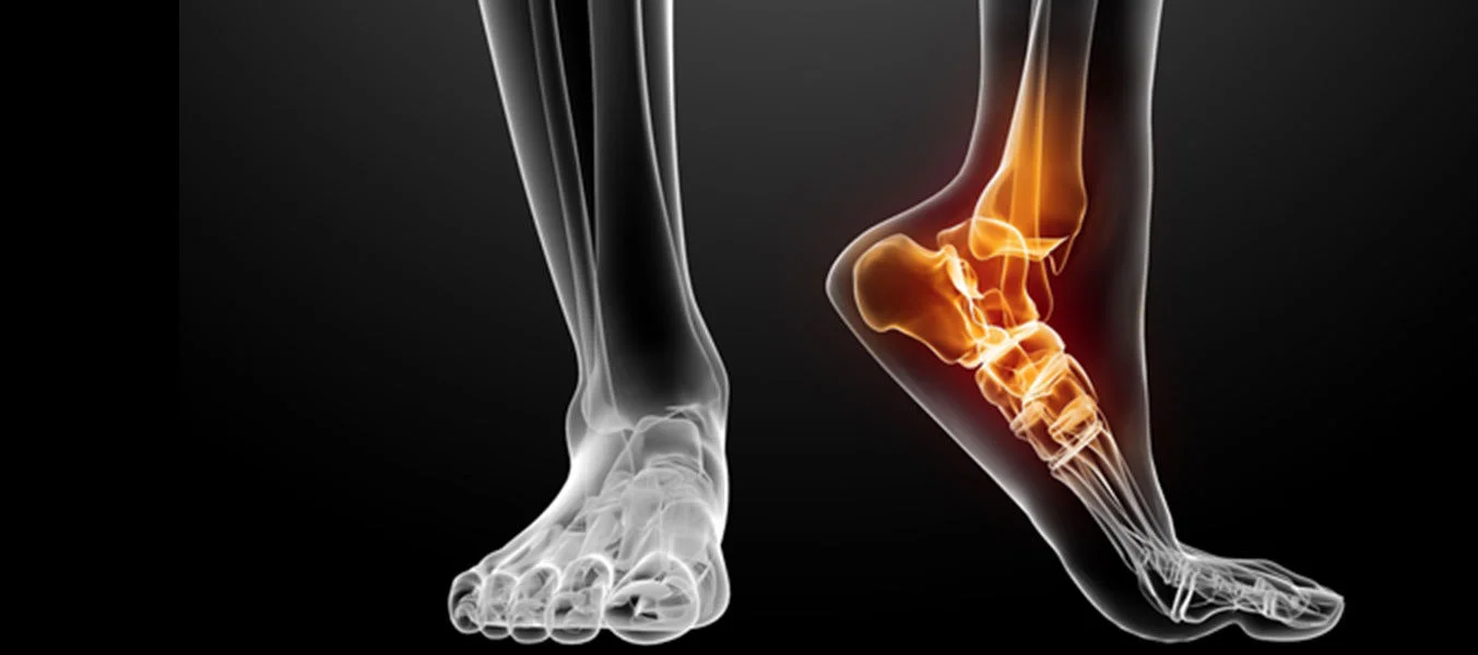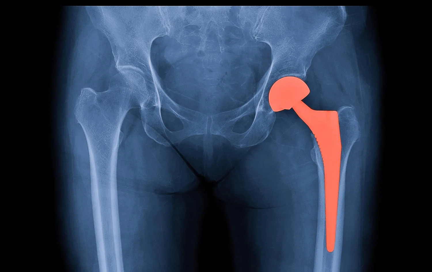
An orthopedic deformity is an abnormality in the alignment, shape, length, rotation, or anatomical relationships of bones and joints.
Deformities may involve the upper or lower limbs, but they can also affect other parts of the skeleton, such as the pelvis and spine.
A deformity may present as:
- angular malalignment,
- rotational abnormality,
- limb-length discrepancy,
- abnormal joint orientation,
- or a combination of these problems.
The goal of deformity correction surgery is not simply to make a bone look straighter. The broader objective is to restore alignment, mechanical axis, rotation, limb length, and joint function as closely as possible to normal.
What Causes Orthopedic Deformities?
Skeletal deformities may be broadly classified as either congenital or acquired.
Congenital Deformities
Congenital deformities develop when the normal formation or growth of the musculoskeletal system is disrupted before birth.
Some are associated with genetic conditions or syndromes, while in many cases no single cause can be identified.
Examples include:
- congenital limb deficiencies,
- developmental abnormalities of the femur or tibia,
- clubfoot,
- congenital hand and foot differences,
- and skeletal dysplasias.
Acquired Deformities
Acquired deformities develop after birth.
Possible causes include:
- fractures that heal in an abnormal position,
- growth-plate injuries,
- bone infections,
- metabolic bone disorders,
- neuromuscular conditions,
- previous surgery,
- bone tumors,
- joint disorders,
- and trauma.
In children, injury to part of a growth plate is particularly important. If one part of the growth plate stops growing while the opposite side continues to grow, progressive angular deformity or limb-length discrepancy may develop.
What Types of Deformities Can Occur?
A deformity does not simply mean that a bone is “bent.” Deformities can be classified as follows:
Angular Deformity
The bone may deviate inward, outward, forward, or backward.
Common terms include:
- varus – the distal part of the limb angles toward the midline,
- valgus – the distal part angles away from the midline,
- procurvatum – forward angulation,
- recurvatum – backward angulation.
Rotational Deformity
A bone may be excessively rotated inward or outward around its longitudinal axis.
Rotational abnormalities may become particularly noticeable during walking, when the feet turn excessively inward or outward.
Limb-Length Discrepancy
One arm or leg may be shorter or longer than the other.
Small differences may cause no significant problem. Larger discrepancies can result in:
- limping,
- pelvic tilt,
- back discomfort,
- abnormal joint loading,
- and functional limitations.
Combined Deformities
Some patients have angular, rotational, and length abnormalities simultaneously.
These multiplanar deformities require more detailed analysis and surgical planning.
Why Should a Deformity Be Treated?
Not every deformity requires surgery.
Treatment should not be based on appearance alone.
Intervention should be considered when the deformity causes:
- pain,
- gait disturbance,
- restricted joint movement,
- limb-length discrepancy,
- loss of function,
- abnormal joint loading,
- or progressive arthritis.
A pronounced alignment abnormality, particularly in the lower extremities, can lead to an abnormal distribution of body weight across the knee, hip, and ankle joints.
Over time, abnormal loading may contribute to secondary problems in adjacent joints.
One of the major goals of deformity correction is therefore to restore more normal load distribution and to protect joint function over the long term.
How Is a Deformity Evaluated?
Accurate analysis and planning are essential.
The assessment should not focus solely on the visibly deformed bone.
In lower-limb deformities, the:
- pelvis,
- hip,
- knee,
- ankle,
- and foot
should be evaluated as a functional unit.
Clinical Examination
Assessment may include:
- limb length,
- joint range of motion,
- muscle strength,
- gait,
- rotational profile,
- joint stability,
- and soft-tissue contractures.
Standing Full-Length Radiographs
Weight-bearing radiographs that show the entire lower limb from the hip to the ankle are particularly important.
They allow measurement of:
- mechanical axis,
- anatomical axis,
- level of deformity,
- magnitude of angulation,
- joint orientation,
- and limb length.
What Is the Mechanical Axis?
The mechanical axis is the line along which body weight is transmitted from the hip to the ankle.
In a normally aligned lower limb, this line passes through a predictable region of the knee.
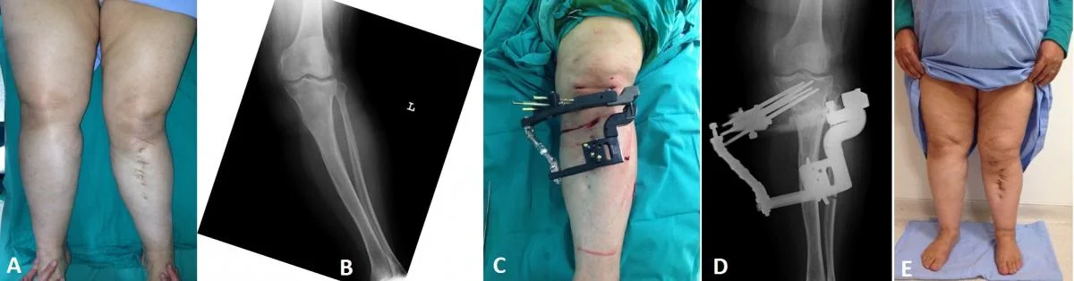
Varus or valgus deformity shifts the mechanical axis, increasing the load on one side of the joint.
A major objective of corrective surgery may therefore be to restore this load-bearing line to a more appropriate position.
Advanced Imaging and 3D Planning
Standard X-rays are not sufficient for every deformity.
When necessary, assessment may include:
- CT,
- MRI,
- rotational measurements,
- and three-dimensional imaging.
For complex multiplanar deformities, computer-assisted planning and 3D models can be especially useful.
CT-based 3D bone models may be used to plan the level and magnitude of an osteotomy before surgery.
In selected patients, patient-specific cutting or positioning guides can also be designed.
Why Is Deformity Treatment Different in Children?
Ongoing growth provides unique treatment opportunities.
In selected pediatric deformities, correction can be achieved without cutting the bone by temporarily influencing one side of the growth plate.
This technique is known as guided growth.
Guided Growth Surgery
Guided growth may be used in children with deformities such as genu valgum or genu varum.
A small plate or similar implant is placed across one side of the growth plate to temporarily slow growth on that side.
The opposite side continues to grow, allowing the deformity to gradually correct.
Timing is crucial because sufficient growth potential must remain.
What Is an Osteotomy?
An osteotomy is a controlled surgical cut in a bone to correct its position.
It is one of the fundamental techniques used in deformity correction.
After the osteotomy, the bone may be stabilized using:
- plates and screws,
- intramedullary nails,
- or external fixation.
Correction may be performed immediately or gradually, depending on the deformity.
Acute Deformity Correction
Some deformities can be corrected completely during a single operation.
The bone is cut, repositioned to the planned alignment, and stabilized with internal fixation.
This is known as acute correction.
Its advantage is that correction is achieved in a single surgical procedure.
However, in large or complex deformities, sudden correction may place excessive tension on nerves, blood vessels, muscles, and other soft tissues. Therefore, acute correction is not suitable for all patients.
Gradual Deformity Correction
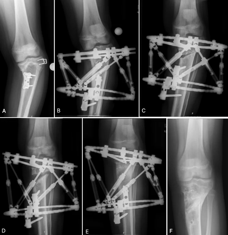
Large or complex deformities can be corrected slowly over days or weeks.
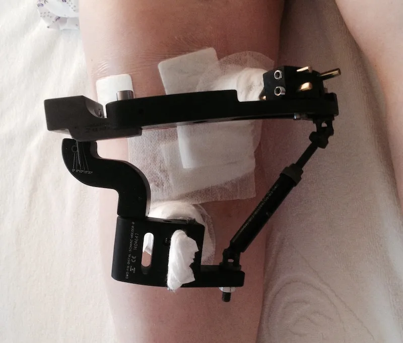
External fixation systems are commonly used for this purpose.
After a controlled osteotomy, small adjustments to the frame are made each day, gradually shifting the bone's position.
Gradual correction may be particularly useful for:
- severe angular deformities,
- rotational abnormalities,
- limb-length discrepancies,
- and combined multiplanar deformities.
The Ilizarov Method and Circular External Fixation
The Ilizarov method is one of the major developments in limb reconstruction and lengthening surgery.
Circular rings are connected to the bone using wires and pins.
The system can be used for:
- deformity correction,
- limb lengthening,
- treatment of nonunion,
- and reconstruction of bone defects.
The method is based on the biological principle of distraction osteogenesis, in which new bone forms as two bone segments are gradually separated under controlled conditions.
Computer-Assisted and Hexapod External Fixation
Modern circular external fixation systems may employ computer-assisted planning.
Hexapod frames can simultaneously correct:
- angulation,
- rotation,
- translation,
- and length.
Radiographic measurements and deformity parameters are entered into specialized software to produce a patient-specific correction program.
The frame is then gradually adjusted according to the prescribed schedule.
Limb-Lengthening Surgery
When one limb is significantly shorter than the other, limb-lengthening surgery may be an option.
The bone is divided in a controlled manner, and the two segments are gradually separated.
New bone forms in the gap between the segments.
This procedure is called distraction osteogenesis.
Traditionally, lengthening has been performed with external fixation.
Today, selected patients may also be treated with motorized or magnetically controlled lengthening intramedullary nails, enabling gradual lengthening without an external frame.
Deformity Correction With Plate and Screw
In suitable cases, an osteotomy can be performed, and the corrected bone can be stabilized with a plate and screws.
This technique is particularly useful when the deformity can be safely corrected in a single stage.
Modern locking plates are available in anatomically contoured designs for many skeletal regions.
Deformity Correction With Intramedullary Nail
Certain long-bone deformities, such as those of the femur and tibia, can be corrected using intramedullary nails placed inside the bone.
One advantage is that the implant remains within the bone rather than outside the limb.
Some modern intramedullary systems can also be used for controlled limb lengthening.
Soft-Tissue Procedures
Not all deformities are caused by bone alone.
Muscles and tendons may develop:
- shortening,
- contracture,
- or imbalance.
Corrective surgery may therefore also include:
- tendon lengthening,
- tendon transfer,
- muscle release,
- or capsular release.
This is particularly important in neuromuscular deformities, where the balance between bone and soft tissue must be considered together.
Is the Goal of Deformity Surgery Always Complete Anatomical Correction?
Not necessarily.
The surgical plan should consider:
- age,
- existing joint damage,
- remaining growth,
- muscle strength,
- neurological status,
- and functional requirements.
In some patients, the most appropriate goal is not a mathematical “zero-degree” correction but rather the best functional and biomechanical alignment for the individual.
What Are the Risks of Deformity Correction Surgery?
Deformity correction procedures range from relatively straightforward to highly complex.
Potential complications include:
- infection,
- nerve or blood vessel injury,
- delayed bone healing,
- nonunion,
- recurrent deformity,
- joint stiffness,
- muscle contracture,
- implant-related problems,
- and pin-site infection when external fixation is used.
Regular follow-up is especially important during gradual correction and lengthening.
The bone is not the only structure adapting during correction; muscles, tendons, nerves, and blood vessels must also adapt to changes in length and alignment.
Rehabilitation After Deformity Surgery
Successful deformity correction depends on more than surgery alone.
Rehabilitation aims to:
- preserve joint motion,
- maintain and rebuild muscle strength,
- prevent contracture,
- progress weight bearing safely,
- retrain gait,
- and restore balance and coordination.
Physical therapy is particularly important during limb lengthening and external fixation treatment.
The Modern Approach to Deformity Correction
Modern deformity correction surgery goes far beyond simply straightening a visibly crooked bone.
Contemporary treatment combines:
clinical examination + biomechanical analysis + precise radiographic measurement + computer-assisted planning + appropriate surgical technique + structured rehabilitation.
The ultimate goal is not merely a straighter-looking limb. It is to create a limb that is:
biomechanically balanced, functional, as pain-free as possible, and capable of protecting the surrounding joints over the long term !!!